8033113536854 - NASTRINO A POI CM.270x1.5 PEZZI 10 - 270x1.5
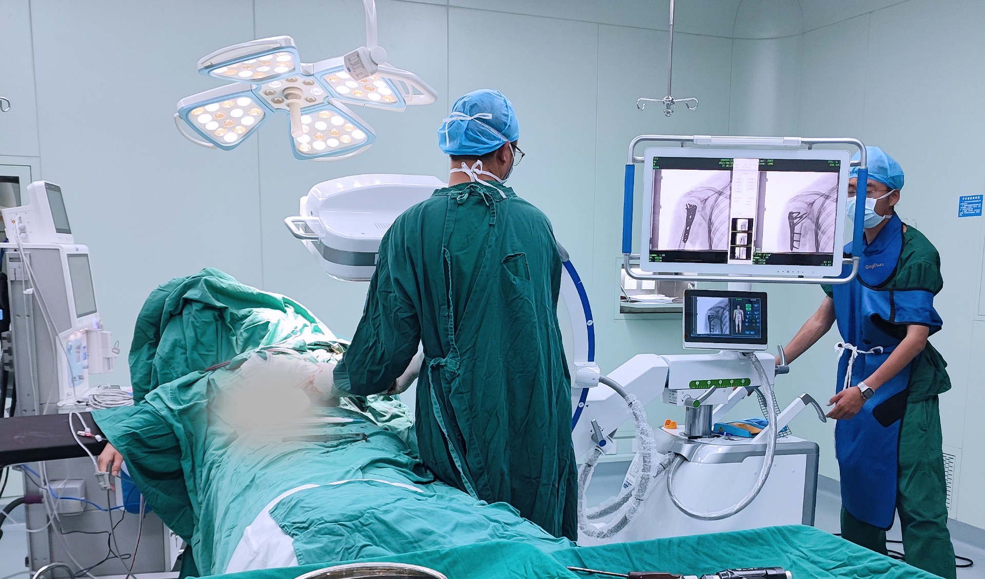
The Large flat-panel all-in-one C-arm helps Pubei County Hospital of Traditional Chinese Medicine to carry out accurate diagnosis and treatment!
Loss of flexibility of positioning the monitor cart wherever needed while positioning the patient and the c-arm to the surgeon’s convenience. Although this may not always be the case. , for instance comes with separate monitor unit.
C-arm machine
For orthopedics, pain management or general surgery 9″ image intensifier would be suitable. Vascular procedures would generally need the 12″ image intensifier. The larger field of view of the 12″ magnification mode enables visualization of larger portion of the body enabling the surgeon to do procedures in a one-shot instead of seeking multiple views as may be the case if a 9” II was used.
A C-Arm machine is an advanced medical imaging device based on X-ray technology. They are primarily used for fluoroscopy capabilities, although they have radiography capability too. C-Arm is called so, due to its C-shaped arm, which is used to connect the x-ray source on one end and the detector on the other.
Radiography involves use of x-ray for imaging and recording the image either digitally or on an x-ray film. Fixed fluoroscopy procedures such as barium meal studies gather real-time moving images using x-ray for visualizing body parts and flow of fluids like blood or contrast agent, to detect blockages etc. Both X-ray (Radiography) and fixed fluoroscopy are used for diagnostic purposes. C-Arm on the other hand, although again a fluoroscopy device, is used to aid surgical procedures.
Rotating anode systems can be used for longer duration and at a higher dose. If in case large number of cases are likely to involve longer scans like run-offs or cross laterals, or scans requiring higher dose for larger patients, the anode is likely to get heated more. In this scenario, the superior heat dispersal of the rotating anode would be preferable option. Popular rotating anode models include the OEC 9600, 9800, Allengers HF 49R, or 59R.
An image intensifier (II) is an integral part of C-Arm machines, converting x-rays into high-intensity light forming brighter images as compared to just a fluorescent screen.
C arm partslabeled
C-Arm machines are widely used during orthopedic, urology, gastroenterology surgeries, other complicated surgical, pain management and emergency procedures, cardiac and angiography studies and in therapeutic studies including stents or needle placements. The Fluoroscopy technology enables high-resolution X-ray images provided in real time, so that the surgeons can do more accurate procedures with better outcomes for the patient. C-Arm systems are also mobile and provide a great deal of flexibility to the surgeon to take images of whole body of the patient from different angles as required.
The choice of type of C-Arm depends on what procedures are going to be done. Full clarity on its proposed use is critical in selection of C-Arm. When buying mobile C-arms, the following main features are key considerations:
A mobile C-arm, comprises of a X-Ray generator, an image intensifier or flat-panel detector and workstation, which has the controls and settings. The C-shaped arm connecting the generator and detector, allows movement horizontally, vertically and around the swivel axes, so that X-ray images of the patient are produced from many angles. The generator emits X-rays that penetrate the patient’s body which is placed in between. The image intensifier or detector converts the X-rays into a visible image displayed on the C-arm monitor. Physician can check anatomical details such as bones, fractures in the bone under consideration vs the position of implants and instruments in real-time, while doing the procedure, without causing damage to other parts.
As is evident from the name – Compact C-Arms come with an all-in-one design where in the monitor; generator, tube, Image intensifier, console and C-arm are all housed in one unit. This compactness saves a lot of space in the operating theatre and removes clutter. Compact systems also cost less than standard-sized systems, while they can handle most common types of cases.

In summary, while compact C-Arms do come with lots of flexibility and lower cost, they may not support wider range of procedures and types of patients. Hence, if we are expecting all types of cases and higher volume of usage, better to go for full-sized C-arm machine.
C-Arm machine with 12″ image intensifiers also tend to be slightly larger in size as a unit, hence they need more space and are also costlier.
3D imaging – C-arm computed tomography is a new and innovative imaging technique. It uses two-dimensional (2D) X-ray projections acquired with a FDP C-arm system to generate images like CT slices, visualized either as cross-sectional images or as 3D data sets using different volume rendering In combination with 2D fluoroscopic or radiographic imaging, 3D C-arm imaging provides valuable information for therapy planning, guidance, and outcome assessment all in the interventional suite.
GE O-arm
We offer all types of imaging equipment including C-Arm machines. We also assist in medical equipment repair & maintenance services at www.Perlove.net, including assistance for AERB licensing.
Most C-arms today come with dual focal spots. However, one may have to choose between stationary or rotating anode options. In the case of a rotating anode tube, the heat of the incoming cathode beam is dispersed evenly across the entire surface of the anode as it rotates. This enables rotating anode users to perform longer scans and at higher doses, without compromising on life of the anode.
Flat panel C-arm
All-in-one compact systems also typically use stationary anode tubes with lower heat capacities, essentially negating the possibility of long-exposure cases..
Higher the generator power, the better it is. But let’s not waste power where it is not necessary. Low-power, mini C-arms are used for instantaneous imaging of extremities such as hands, feet, wrists, elbows, ankles etc. both in out-patient settings as well as surgeries, such as during external reduction and fixation of fractures. C-arms with higher power are necessary in surgical procedures. The benefit of increased x-ray power is that it allows greater flexibility for imaging with shorter exposure times and reducing the risk for error. These benefits may be very important for pediatric and hig
What is a 3D C-arm
Copyright © - Nanjing Perlove Medical Equipment Co., Ltd. All Rights Reserved Internet Drug Information Service Qualification Certificate [(Su) - Non-operational -2023-0073] Company address: No. 97/99, Wangxi Road, Jiangning District,Nanjing,China

Flat-panel detectors (FDP) are increasingly replacing image intensifiers (II) on mobile C-arm systems. The advantages of a flat panel detector include: lower patient dose and increased image quality. There is no deterioration of the image quality over time. With FDP, the overall physical size of the C-arm is also reduced significantly as compared to usage of image intensifier.
On the other hand, if mainly short, lower-dose studies, such as pain management needle placement or hand and foot specialties, a stationary anode tube would suffice. Models like the Siemens Compact L, Allengers HF 49, Philips Surgico 60-D HF, Siemens Multimobil 5C, RMS Corus C-arm are popular models with stationary anode.
Mobile C-arm units should be maneuverable around hospitals and provide positioning flexibility. The dimensions of the unit are therefore important. A great C-arm depth may be preferable, but it should not be too large making it difficult to move around in the OT. For example, it must be deep enough to accommodate all sizes of patients and low enough to fit underneath the hospital’s beds and operating theatre tables. Hence from the surgeon’s point of view – Orbital, horizontal, vertical and lateral movements, angulation and swivel range are all important considerations. X-ray source – image intensifier distance with isocentric rotation (where center of rotation is the same as the midpoint between the x-ray tube focal spot and the image intensifier), is also an important consideration.
Image intensifiers come in a range of input field of view (FOV) diameters for diagnostic imaging applications, from 6 inches (15 cm FOV) to 16 inches (40 cm FOV), and many dimensions in between, depending on the type of imaging procedure, the C-arm is required for. While the higher II size/diameter provides with larger field of view, the lower II mode gives greater degree of magnification and clarity.

Most C-Arm machines today come with high resolution CCD camera, two 17” or bigger monitors for display, storage of atleast 100 frames or more, as well as USB data port for transfer of images. Older systems like Siemens Multimobile 5C had only standard four frame image memory in addition to Last Image Held. It is important to check the image handling capability of the C-arm depending on the way it will be used. If the C-Arm is likely to be used only during the procedure, with no need for future referral to the images or transfer and storage in a Hospital Information System or Electronics medical records of the patient, then one need not worry about the storage and transfer capacity.
C-arms come with single-field, dual-field or triple-field image intensifiers (II). So, in a triple-field II, the 9″ image intensifiers could come in 4.5″, 6″ and 9″ size while the 12″ image intensifier could be in the 6″, 9″ and 12″ size. Older models like Siemens Multimobile 5C came with 6” and 9” dual modes.


 Neil
Neil 
 Neil
Neil